Medical AI Platform
プログラミング不要な
医用AI開発・解析
プラットフォーム
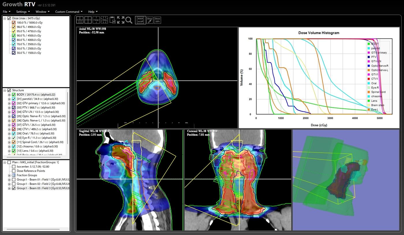
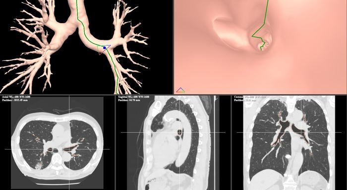
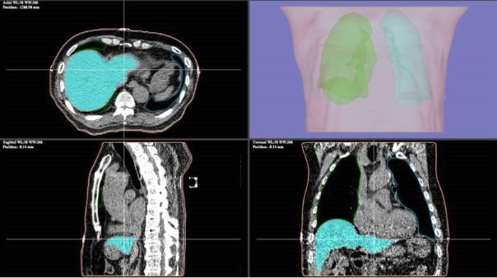
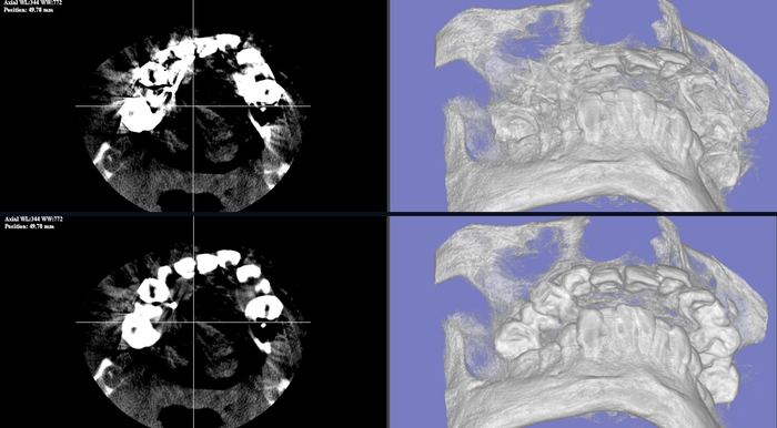
Growth RTV シリーズは、3D医用画像(DICOM)のAI訓練・臓器自動抽出をノーコードで実現する医用AI開発プラットフォームです。研究・教育向け無償版(Growth RTV Cloud)から幅広く展開しています。
NVIDIA
Inception Partner
NVIDIA MONAI対応
SCROLL